

Introduction
Low-dose CT scans and magnetic resonance imaging (MRI) using T2- and T1-weighted contrasted enhanced images are the standard methods of bone staging for newly diagnosed and relapsed multiple myeloma (MM) patients. Fluoro-desoxy-glucose positron emission tomography (18F-FDG PET) is also a validated procedure for the imaging of MM bone lesions and the evaluation of extramedullary plasmacytoma. Furthermore, PET CT is useful to determine the prognosis and to assess the minimal residual disease. A new hybrid technology combining PET and MRI provides metabolic and anatomic information simultaneously. We evaluated the results obtained using this new examination.
Materials and Methods
Thirty-nine MM patients were prospectively included in this single center observational study. Fifty-one PET-MRI scans were performed in 4 patient populations: 1/ newly diagnosed smoldering MM (NDSMM, n =10); 2/ newly diagnosed symptomatic MM (NDMM, n=18); 3/ MM patients with long term follow-up (FU, n=17); 4/ MM patients with biochemical relapse (Rel, n=6). The imaging protocol consisted of T1-weighted, DIXON and diffusion-weighted (b=50 and b=800) axial sequences from the head to the knees simultaneously acquired by PET, followed by coronal and sagittal T1-weighted and T2-weighted sequences centered on the full spine and basin. MRI was considered to be positive in the case of diffuse bone marrow infiltration (DI), diffuse patchy bone marrow infiltration (DPI), focal bone marrow lesions (FL) or extra-medullary disease (EMD). PET was considered to be positive when FDG-avid focal bone lesions, a diffuse medullary area and/or FDG-avid EMD were observed. Medullary compression was also evaluated using MRI.
Results
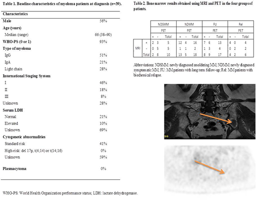
The characteristics of the patients are summarized in Table 1. They were classified according to the ISS scale (I, n=18; II, n=7; III, n=3; missing, n=11) and none had high-risk cytogenetics or extra-medullary disease. The hybrid PET-MRI technique provided good image quality in all cases with no artefacts. No medullary compression was detected by MRI. Among the overall 51 PET-MRI examinations, 38 (74%) were positive for MRI and 27 (53%) for PET. Table 2 summarizes the bone marrow results obtained using MRI and PET in the four groups of patients. EMD was not detected by either MRI or PET. In NDSMM patients, MRI was positive and PET was negative in 30%, while none were positive only for PET. In NDMM, MRI was positive and PET was negative in 22% of cases, while 5% were positive only for PET (Figure 1 shows a negative PET and a positive MRI scan in the same patient). In FU patients, 35% of MRI scans remained positive with PET negative, while 6% were PET-positive and MRI-negative. In the case of relapsed patients, MRI and PET scans were in 100% agreement.
Conclusions
Using PET-MRI, in addition to a search for medullary compression, MRI can provide additional bone marrow information with respect to PET in about 22 to 30% of cases, particularly in NDSMM and NDMM. In a single examination, PET-MRI can detect bone, as well as potential extra-medullary lesions and medullary compression, without irradiation. The baseline PET examination is useful to evaluate the prognosis.
Legend to Figure 1: PET and MRI scans in an asymptomatic myeloma patient. PET is negative but MRI reveals a bone lesion in the left iliac crest (arrow).
Leblond:Janssen: Honoraria; Lilly: Honoraria; Astra Zeneca: Honoraria; Roche: Honoraria; Amgen: Honoraria; Gilead: Honoraria; Beigene: Honoraria; Beigene: Membership on an entity's Board of Directors or advisory committees; Gilead: Membership on an entity's Board of Directors or advisory committees; AbbVie: Membership on an entity's Board of Directors or advisory committees; Astra Zeneca: Membership on an entity's Board of Directors or advisory committees; Janssen: Membership on an entity's Board of Directors or advisory committees; AbbVie: Honoraria.

This feature is available to Subscribers Only
Sign In or Create an Account Close Modal